
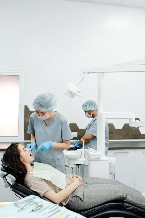
Most children have their first dental radiographs between the ages of five and six, although the honest clinical answer is that there is no fixed age at all. Radiographs are taken when there is a specific diagnostic reason, such as back teeth that touch and cannot be inspected visually, a high risk of decay, delayed eruption, or a history of trauma. This is a routine, low risk part of preventive care rather than anything alarming, and a gentle first visit at Honey Smile Dentistry will establish whether imaging is needed yet or can safely wait.
The guiding principle used worldwide is selection criteria, meaning images are prescribed individually after an examination. Blanket screening of every child on a calendar schedule is not considered good practice.
Choosing a Dental Clinic in Toronto equipped with modern digital sensors matters, because digital systems cut radiation dose dramatically compared with the film used decades ago.
For adults, imaging plays a very different role, and three dimensional scanning is essential when planning dental implants in Toronto and North York so bone volume and nerve position are known before surgery.
The same diagnostic detail supports predictable cosmetic dentistry in Toronto, where root position and bone contour influence the final appearance of a smile.
If your child has never been examined, book a friendly introductory visit first. You can request an appointment online or call +1 437-525-2715 and mention that it is a first appointment.
Direct Answer: Typical Timing by Age
- Ages one to three: a first dental visit is recommended by the first birthday or within six months of the first tooth appearing. Radiographs are rarely needed unless there is trauma or visible decay.
- Ages four to six: the usual window for the first images, most often two small bitewing films, once the back baby teeth have closed contacts.
- Ages six to eight: a panoramic image may be taken to check the number and position of developing permanent teeth.
- Ages seven to nine: an early orthodontic assessment often includes imaging to identify crowding, impaction or missing teeth.
- Adolescence: wisdom tooth development is reviewed, usually from around age sixteen onwards.
Why Radiographs Are Prescribed, Not Routine
A dental radiograph answers a question that cannot be answered by looking. Nearly half of decay between back teeth is invisible to the naked eye until it is advanced, and early lesions found on a bitewing image can often be arrested with fluoride rather than a filling.
Clinical indications include:
- Back teeth in contact, where the surfaces between them cannot be seen.
- A previous history of cavities or visible white spot lesions.
- Unexplained pain, swelling or discolouration of a tooth.
- Trauma to the face or teeth.
- A tooth that has not erupted when expected, or teeth erupting in an unusual position.
- Suspected extra teeth, missing teeth or abnormal development.
- Planning for orthodontics, extractions or restorative treatment.
How Safe Are Dental X-Rays for Children?
Modern dental radiography uses a very small radiation dose. A single digital bitewing delivers a fraction of the natural background radiation a person receives from the environment over a normal day. Protective measures reduce exposure further.
Safety measures a good practice uses
- Justification. Every image must have a clinical reason recorded in the notes.
- Digital sensors. These require far less radiation than traditional film.
- Rectangular collimation. Narrowing the beam to the sensor size reduces scattered radiation significantly.
- Child specific settings. Exposure time and sensor size are adjusted for smaller anatomy.
- Thyroid protection. A collar is used where it does not obscure the area being imaged.
- Fastest possible technique. Careful positioning avoids repeat exposures.
Radiographs are never taken as a routine formality. A Top Dentist in Toronto follows the principle of keeping doses as low as reasonably achievable while still obtaining the diagnostic information required to protect the child.
Comparison: Types of Dental Image
- Bitewing: shows the crowns of upper and lower back teeth together. Best for detecting decay between teeth and checking bone levels.
- Periapical: shows a whole tooth including the root and surrounding bone. Used for pain, trauma and root assessment.
- Panoramic: a single broad view of both jaws. Useful for development, wisdom teeth and jaw pathology, but not detailed enough for early decay.
- Cephalometric: a side profile view used in orthodontic planning.
- Cone beam CT: three dimensional imaging reserved for complex cases such as implant planning or impacted teeth, because the dose is higher than a standard film.
How Often Should Images Be Repeated?
Intervals depend on decay risk rather than the calendar. Common guidance is:
- Higher risk children: bitewings roughly every six to twelve months while risk remains elevated.
- Lower risk children: every twelve to twenty four months.
- Lower risk adults: every two to three years.
- Patients with active periodontal disease: imaging as clinically required to monitor bone levels.
Risk is reassessed at every check up, so an interval that was appropriate last year may lengthen or shorten. This individualised approach is a hallmark of a Best Dental Clinic in Toronto standard of care.
Preparing a Child for Their First X-Ray
- Use simple, positive language. Describe it as a photograph of the teeth. Avoid words such as pain, needle or shot.
- Book at a good time of day. Mornings usually work better than the end of a long day.
- Let the team lead. Clinicians experienced with children use tell, show, do techniques that build trust quickly.
- Stay calm yourself. Children read parental anxiety instantly.
- Practise at home. Holding still and keeping the mouth closed on a small sensor for a few seconds is easy to rehearse.
- Praise afterwards. A positive first experience shapes attitudes to dental care for years.
Myths That Worry Parents Unnecessarily
Myth: children should never have x-rays
Avoiding necessary imaging can allow decay to progress silently into the nerve, leading to infection, pain and treatment that is far more invasive than a small filling would have been.
Myth: baby teeth do not matter
Primary teeth guide the eruption of permanent teeth, support speech development and chewing, and infection in a baby tooth can damage the permanent tooth forming beneath it.
Myth: dental treatment for children is frightening and painful
Behaviour guidance, topical anaesthetic and unhurried appointments make paediatric care comfortable. Most children who are introduced gradually cope extremely well, and anxiety is usually inherited from adults rather than from experience.
Myth: one x-ray is enough for years
Images capture a moment in time. Decay and development change, so review intervals exist for good reason.
Family Focused Care in North York
Honey Smile Dentistry is a premium boutique Dental Office in North York at 12 Inn on the Park Dr, Toronto, ON, Canada, M3C 0P9. The practice combines advanced digital diagnostic technology with a warm, sophisticated environment where children and nervous adults are given time to settle.
Preventive care, cosmetic dentistry, dental implants and teeth whitening are all available, so families can stay with one team as needs change over the years. Parents searching for a dependable Family Dentist in Toronto or the Best Dentist in North York often choose the clinic specifically for its patient first, comfort led approach.
The team is accepting new patients of all ages. Email info@honeysmiledentistry.ca, call +1 437-525-2715, or book online in moments, with same day appointments offered where possible. Urgent problems such as a knocked out tooth after a playground accident are treated as Emergency Dental Clinic in Toronto priorities.
Frequently Asked Questions
Can my child refuse to have an x-ray taken?
Yes, and a good clinician will not force the issue. Alternative approaches include a smaller sensor, a different film position, or delaying the image and monitoring clinically until the child is ready.
Are dental x-rays safe during pregnancy?
Necessary dental radiographs with appropriate shielding are considered safe at any stage of pregnancy, and untreated dental infection carries its own risks. Always tell your dental team that you are pregnant so they can adjust the plan.
How much radiation is in a single dental x-ray?
A digital bitewing delivers a very small dose, comparable to a short period of everyday background radiation from the environment. Your dental team can provide precise figures for the equipment they use.
Will my child need x-rays at every check up?
No. Imaging is prescribed according to individual decay risk and clinical need, and children with healthy teeth and low risk may go two years or longer between images.
What happens if an x-ray shows early decay?
Early lesions confined to enamel can often be arrested with fluoride varnish, dietary advice, improved brushing and close monitoring, without any drilling at all. Finding decay early is exactly what makes conservative treatment possible.
Conclusion
Most children have their first dental radiographs around age five or six, but the real trigger is clinical need rather than a birthday. Modern digital imaging uses a very low dose, and finding decay early usually means simpler, gentler treatment. Ask your dental team to explain the reason for each image, and keep review intervals matched to your child individual risk.
This article offers general guidance only and does not replace an individual clinical examination or professional dental advice.